Disaster Strikes: A Young Father's Life Hangs by a Thread
On April 4, 2026, a 28-year-old man was rushed to Chongqing HYGEIA Hospital's emergency department following a severe traffic accident. Upon admission, he was in deep coma and unresponsive to verbal stimuli. Neurological examination revealed critical signs: bilateral pupils showed no light reflex whatsoever — the classic clinical presentation of late-stage brain herniation, a near-fatal condition.
Emergency head CT imaging revealed a devastating picture: a large crescent-shaped mixed-density hematoma under the right fronto-temporo-parietal skull, measuring up to 19mm at its thickest point. Brain tissue and the lateral ventricle were severely compressed, with the midline structure shifted 14mm to the left. The diagnosis was clear: massive right fronto-temporo-parietal subdural hematoma with both subfalcine herniation and transtentorial herniation — dual brain herniation, one of the most lethal conditions in neurosurgery.
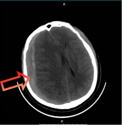
Pre-operative head CT: Massive right fronto-temporo-parietal subdural hematoma (up to 19mm thick), with severe brain compression and 14mm midline shift — dual brain herniation in progress.
The hospital immediately activated the severe traumatic brain injury emergency green channel, mobilizing all relevant departments into emergency preparedness.
Extreme Speed: Emergency Craniotomy in Under 1 Hour
Time is brain — every minute of delay exponentially increases the risk of irreversible brain tissue damage. From the patient's admission to the start of surgery, the entire process took less than 1 hour, intercepting the fatal progression before brain tissue necrosis became irreversible.
That same day, the emergency surgical team performed a highly complex and high-risk procedure: right fronto-temporo-parietal subdural hematoma evacuation + decompressive craniectomy. This operation offers virtually zero margin for error. In a state of brain herniation, the patient's brain tissue is severely swollen and extraordinarily fragile — akin to performing surgery on soft tofu, where the slightest touch could cause rupture. During the procedure, acute brain bulging, cardiac arrest, or massive hemorrhage could occur at any moment, placing extreme demands on the surgeon's precision, emergency response capability, and anatomical mastery.
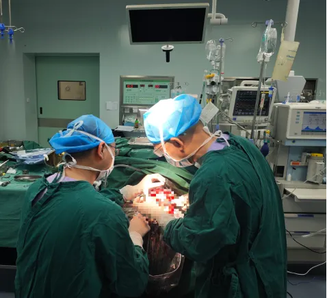
The neurosurgery team at Chongqing HYGEIA Hospital operating room: Dr. Tong Jianguo and Dr. Huang Shaokuan leading the emergency craniotomy with precision and speed.
Drs. Tong Jianguo and Huang Shaokuan of Chongqing HYGEIA Hospital led the team with rapid response, calm composure, and meticulous precision. They thoroughly evacuated the intracranial hematoma, fully decompressed the compressed brain tissue, and completed a standard large decompressive craniectomy — completely relieving the life-threatening pressure on the brain and fundamentally reversing the brain herniation crisis.
Postoperative Milestone: Brain Herniation Crisis Reversed
After surgery, the patient's bilateral pupils significantly constricted and light reflexes became elicitable — a clear sign that the fatal brain herniation compression had been completely relieved.
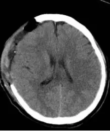
Post-operative CT: Intracranial hematoma completely evacuated, midline structures returned to normal position — brain herniation fully resolved.
Two Months of Multidisciplinary Battle: From Coma to Recovery
The patient was transferred to the ICU for advanced life support and multidisciplinary collaborative treatment. Although the intracranial hematoma had been completely evacuated and the brain herniation promptly and effectively resolved, the patient remained critically ill. The postoperative period presented multiple complications: recurrent high fever and mixed multi-pathogen infections created a series of life-threatening challenges.
Through multidisciplinary joint treatment, the medical team adhered to the philosophy of early rehabilitation and full-course intervention. They immediately coordinated with the rehabilitation department to develop personalized rehabilitation plans for limb function, neurological recovery, and pulmonary function. This approach maximized the reduction of sequelae and promoted functional recovery.
After nearly two months of treatment, the patient successfully had the tracheostomy tube removed, achieving complete airway closure and full recovery of spontaneous breathing. Currently, the patient is conscious and clear, fluent in conversation, with completely normal cognitive function, able to get out of bed and move independently, and capable of living independently.
Not Just a Medical Victory — A Family Saved
This hospital-wide multidisciplinary mobilization at Chongqing HYGEIA Hospital not only demonstrated the hospital's硬核 capabilities in ultra-rapid emergency response for severe traumatic brain injury and stepwise precision treatment for complex critical infections, but also reflected rigorous teamwork and the unwavering commitment of medical professionals who never give up. For one life, for one family, they wrote a new chapter.
Expert Team
Dr. Tong Jianguo
Director of Neurosurgery, Chongqing HYGEIA Hospital
Leading expert in neurosurgical emergency and trauma surgery. Specializes in emergency craniotomy for severe traumatic brain injury, brain tumor microsurgery, and cerebrovascular disease intervention. Dr. Tong has extensive experience in high-risk neurosurgical procedures under critical conditions.
Dr. Huang Shaokuan
Neurosurgery Specialist, Chongqing HYGEIA Hospital
Experienced neurosurgeon specializing in emergency trauma surgery, intracranial hematoma evacuation, and decompressive craniectomy. Dr. Huang brings steady hands and calm judgment to high-pressure emergency neurosurgical procedures.
Media Coverage
This case has been widely reported by mainstream media in Chongqing:
- Chongqing Voice Radio (重庆之声) — Featured coverage of the hospital's emergency response capabilities
- Chongqing Eye (第1眼) — In-depth report on the multidisciplinary rescue and patient recovery journey
Emergency Neurosurgery in China?
Contact HYGEIA Healthcare's international patient team to learn about our trauma surgery capabilities, emergency response protocols, and multidisciplinary critical care expertise across our hospital network.
Book a Consultation— Chongqing HYGEIA Hospital, Brand & Communications Department —
← Back to News