The Crisis: A Perfect Storm of Risk Factors
The patient had undergone right-sided modified radical mastectomy with bilateral breast reconstruction using implants for breast cancer. Even before surgery, she was in a hypercoagulable state — placing her firmly in the high-risk category for venous thromboembolism. On the evening of post-operative day 4, disaster struck without warning.
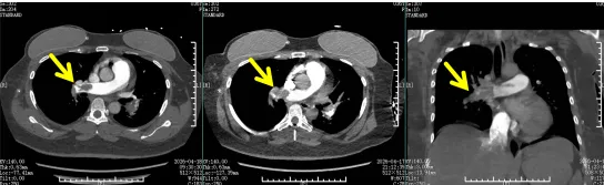
She suddenly developed severe dyspnea, palpitations, profuse sweating, and pain in both lower extremities. Her blood pressure plummeted. The breast surgery team immediately recognized the red flags and rapidly completed bedside arterial blood gas analysis and emergency CTPA (CT pulmonary angiography). The results were alarming:
The main pulmonary arteries and multiple branches on both sides were packed with large clots. The right pulmonary artery was nearly completely occluded. Cardiac arrest could occur at any moment.
Pre-intervention CTPA: Massive thrombi filling the main and segmental pulmonary arteries bilaterally, with near-complete occlusion of the right pulmonary artery — a life-threatening condition requiring immediate intervention.
The Dilemma: Bleeding Risk vs. Clot Crisis
The challenge was not simply removing the clot — it was doing so safely. The patient had undergone major oncological surgery only days earlier. Her chest wall and bilateral breast surgical wounds were still healing. Two standard treatments were effectively off the table:
- Systemic thrombolysis (IV clot-busting drugs) would almost certainly trigger fatal hemorrhage from the fresh surgical sites.
- Open surgical embolectomy (thoracic surgery to remove clots) carried prohibitive trauma — the patient's body could not tolerate a second major operation so soon after mastectomy.
The breast surgery team immediately activated the hospital-wide emergency response, calling for urgent consultation from Interventional Radiology, ICU, and Medical Imaging. Within minutes, multidisciplinary experts assembled at the bedside.
The Solution: A Three-Pronged Minimally Invasive Strategy
Dr. Gan Dengwei, Deputy Director of Interventional Radiology, proposed an innovative one-stop minimally invasive approach designed specifically for this high-risk scenario:
- Mechanical aspiration thrombectomy — A catheter inserted via the femoral vein navigates directly into the pulmonary arteries to physically extract clot fragments under negative pressure.
- Local targeted thrombolysis — A fraction of the systemic dose of thrombolytic drug is infused directly at the clot site, maximizing clot-dissolving effect while minimizing systemic bleeding risk.
- Inferior vena cava (IVC) filter placement — A filter is deployed in the vena cava to trap any remaining lower-extremity clots before they can reach the lungs, preventing recurrent pulmonary embolism at its source.
This strategy addressed all three critical objectives simultaneously: rapid obstruction relief, minimal bleeding exposure, and secondary prevention.
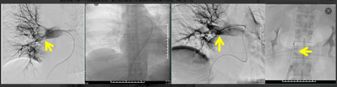
Intra-procedural DSA imaging: Thrombus aspiration catheter positioned in the pulmonary arterial tree. Dark-red thrombus fragments are being mechanically extracted under real-time fluoroscopic guidance (yellow arrows indicate catheter position and residual thrombus).
The Midnight Procedure: Precision Under Pressure
At 23:50, the procedure began. Under real-time DSA (digital subtraction angiography) navigation, the aspiration catheter advanced from the femoral vein upward, precisely reaching the occluded segments of both pulmonary arteries.
Through repeated cycles of negative-pressure aspiration, dark-red thrombus fragments were gradually withdrawn. Angiography showed pulmonary arterial flow progressively restoring, and blood oxygen saturation began climbing. The team then administered localized thrombolytic infusion directly into the occluded segments — using only a fraction of the systemic drug dose — efficiently dissolving residual thrombus while completely avoiding the bleeding risk posed by the patient's unhealed surgical wounds.
Upon completion of aspiration and local thrombolysis, an IVC filter was deployed in the inferior vena cava — functioning as an interception net that firmly traps any dislodged lower-extremity clots, eliminating the root cause of recurrent pulmonary embolism.
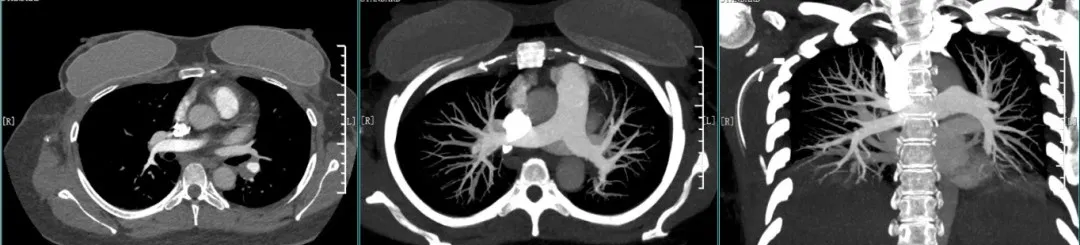
Post-intervention CT: Pulmonary arterial patency markedly improved with significant reduction in thrombus burden (yellow arrows show comparison areas), confirming successful mechanical aspiration and targeted thrombolysis.
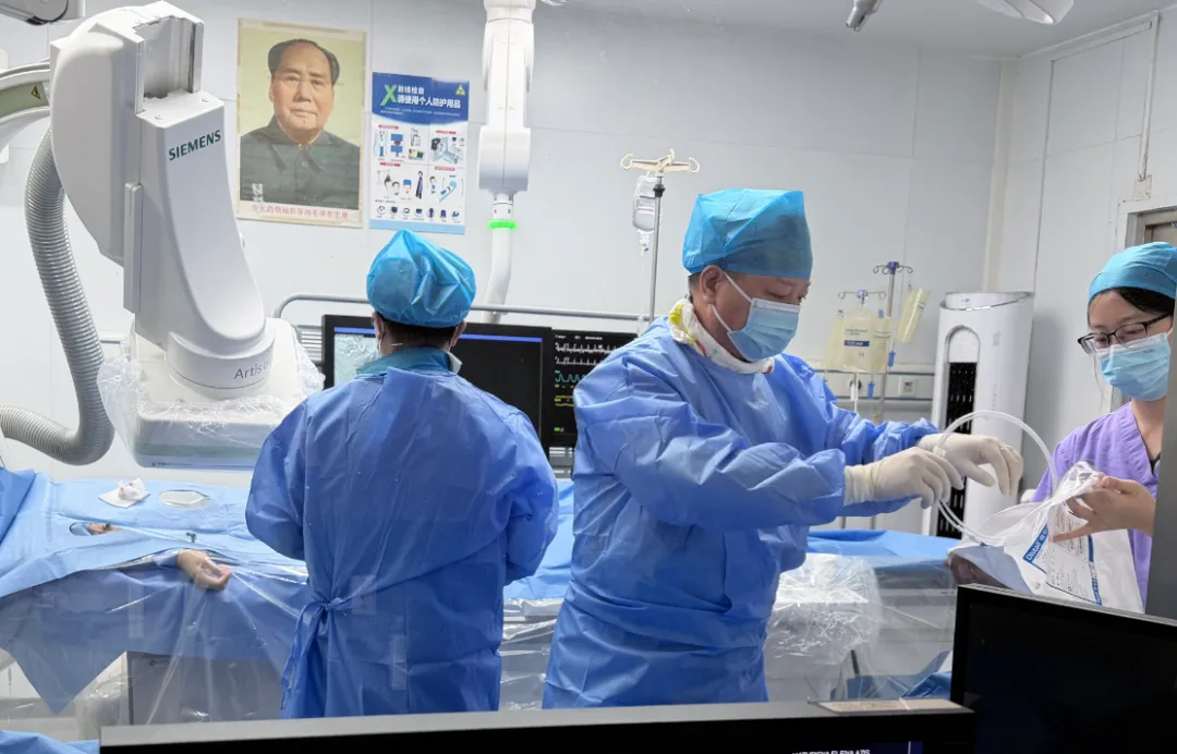
The interventional radiology team at work in the DSA catheterization laboratory (SIEMENS Artis system) at Chongqing HYGEIA Hospital, performing the life-saving pulmonary embolism intervention.
Rapid Recovery: From Critical to Discharged
The entire procedure lasted under two hours. Upon completion:
- Blood oxygen saturation rose from 80% (pre-op) to 97%
- Blood pressure stabilized
- Acute dyspnea and tachypnea resolved
The patient was transferred to ICU for close monitoring. By the following day, arterial blood gas analysis showed oxygen partial pressure recovered to 152 mmHg, and lactic acid levels returned to normal range — clear indicators of successful tissue reperfusion.
She continued on anticoagulation therapy after discharge. One month later, follow-up CTPA confirmed that bilateral pulmonary artery thrombi had essentially disappeared. The patient recovered smoothly and was discharged home.
A Model of Multidisciplinary Excellence in Critical Care
This successful rescue demonstrates the power of tightly coordinated multidisciplinary collaboration at Chongqing HYGEIA Hospital. The breast surgery team's rapid recognition, medical imaging's precise localization, the interventional team's minimally invasive expertise, and ICU's comprehensive perioperative support formed an unbroken chain of care. For post-oncological-surgery patients facing the dual threat of thrombotic complication and bleeding contraindication, this "mechanical aspiration + local thrombolysis + IVC filter" one-stop interventional protocol offers a proven lifeline. Chongqing HYGEIA Hospital continues to refine its full-spectrum diagnostic and therapeutic capabilities, building a robust defense line for oncology and post-surgical patients through technical excellence and seamless multidisciplinary coordination.
Expert Team
Vice President Zhou Peihua
Vice President & MDT Team Leader, Chongqing HYGEIA Hospital
Led the hospital-wide emergency MDT response, coordinating Interventional Radiology, ICU, Breast Surgery, and Medical Imaging teams. Oversaw clinical decision-making and procedural planning for this high-complexity case involving competing risks of hemorrhage and thrombosis.
Dr. Gan Dengwei
Deputy Director of Interventional Radiology, Chongqing HYGEIA Hospital
Designed and executed the three-pronged minimally invasive intervention strategy: mechanical aspiration thrombectomy, local targeted thrombolysis, and IVC filter placement. Dr. Gan specializes in endovascular procedures for acute vascular emergencies and has extensive experience in managing high-bleeding-risk patients through targeted interventional techniques.
Media Coverage
This case has been reported by mainstream media in Chongqing:
- Chongqing Voice Radio (重庆之声) — Featured coverage highlighting the hospital's emergency interventional capabilities
- Chongqing Eye / Di Yi Yan (第1眼) — In-depth report on the multidisciplinary rescue process and patient recovery journey
Need Emergency Vascular or Oncology Care in China?
Contact HYGEIA Healthcare's international patient team to learn about our interventional radiology capabilities, MDT emergency protocols, and comprehensive care for complex post-surgical complications across our nationwide hospital network.
Book a Consultation— Chongqing HYGEIA Hospital, Brand & Communications Department —
← Back to News